
The Complete Guide to the Echocardiogram Procedure: What Patients and Clinicians Should Know
Explore the comprehensive aspects of the echocardiogram procedure, including types, detailed steps, interpretations, and technological advances. This article helps patients and clinicians understand the importance of echocardiography in diagnosing and managing heart conditions, emphasizing its safety, versatility, and evolving innovations.
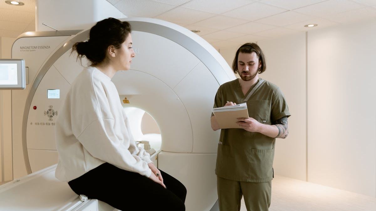
Introduction: Why an Echocardiogram Procedure is a Vital Diagnostic Tool
In contemporary cardiology, the echocardiogram procedure has become an essential, non-invasive imaging technique that enables clinicians to assess heart health with remarkable detail. It provides critical insights into heart function, structural abnormalities, and blood flow, guiding diagnosis, treatment, and ongoing management of cardiovascular diseases. For patients, understanding this procedure demystifies the process and fosters cooperation that leads to more accurate and informative results.
Different Types of Echocardiogram Procedures and Their Clinical Indications
1. Transthoracic Echocardiogram (TTE)
This is the most widely performed echocardiogram, offering a window into the heart using a transducer placed on the chest wall. Especially useful for detecting overall heart size, ejection fraction, valve function, and pericardial effusion, TTE is often the first-line diagnostic test for suspected heart disease.
2. Transesophageal Echocardiogram (TEE)
When higher resolution images are required, especially for detecting small thrombi or infection in the heart valves, TEE becomes invaluable. It involves inserting a flexible probe into the esophagus, providing clearer views of posterior heart structures, atria, and aorta. TEE’s utility extends to pre-surgical planning and intraoperative guidance.
3. Stress Echocardiogram
This procedure combines echocardiography with physical or pharmacologic stress to evaluate how the heart responds under exertion. It helps in diagnosing ischemia, assessing functional capacity, and planning interventions such as coronary artery bypass grafting or stenting.
Step-By-Step: Conducting a Typical Echocardiogram Procedure
Pre-Procedure Preparation
Patients are advised to wear comfortable clothing and avoid caffeine or certain medications, based on physician instructions. If sedation or TEE is planned, fasting may be required. A detailed history helps tailor the procedure to individual needs.
The Actual Procedure
The standard transthoracic echocardiogram involves applying conductive gel on the chest and moving the transducer across different positions to obtain multiple views. During TEE, sedation is administered, and the transducer is guided into the esophagus under monitoring. For stress tests, patients may walk on a treadmill or receive medication to simulate exertion.
Post-Procedure Follow-Up
Most patients can resume normal activities immediately after a TTE. Following TEE or pharmacologic stress echocardiogram, recovery may include observation for side effects like sore throat or side effects from medication. The cardiologist reviews images and discusses findings with the patient.
Interpreting Results and Their Impact on Patient Care
Results from an echocardiogram procedure inform diagnosis, monitor disease progression, and evaluate response to treatment. For instance, detecting decreased ejection fraction can prompt initiation of heart failure therapy, while identifying valvular abnormalities may lead to surgical intervention.
Advantages of the Echocardiogram Procedure
Its non-invasive nature, real-time imaging, and ability to assess multiple aspects of cardiac function make the echocardiogram procedure indispensable. Technological advancements like 3D imaging have further improved diagnostic accuracy and procedural efficiency.
Patient Experiences and Case Studies
Many patients report feeling reassured after undergoing an echocardiogram, especially when results show normal heart function. In complex cases, early detection through echocardiography has led to timely interventions, significantly improving outcomes. Recent case studies demonstrate its role in diagnosing rare congenital defects and guiding minimally invasive procedures.
Future Directions and Innovations in Echocardiography
The ongoing development of high-resolution 3D echocardiography, contrast-enhanced imaging, and portable devices is transforming the landscape of cardiac diagnostics. Experts advocate for wider adoption of these innovations to facilitate earlier detection and personalized treatment strategies.
Conclusion
The echocardiogram procedure represents a cornerstone of modern cardiology, offering detailed insights into heart health with safety and efficiency. Educating patients about the process enhances cooperation and results in better care. As technology evolves, the role of echocardiography will undoubtedly expand, further empowering clinicians and patients alike.